3d Tee Mitral Valve Anatomy
The standard modalities of real time 3d tee have recently been described 3.

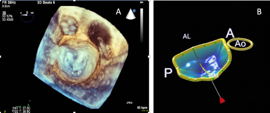
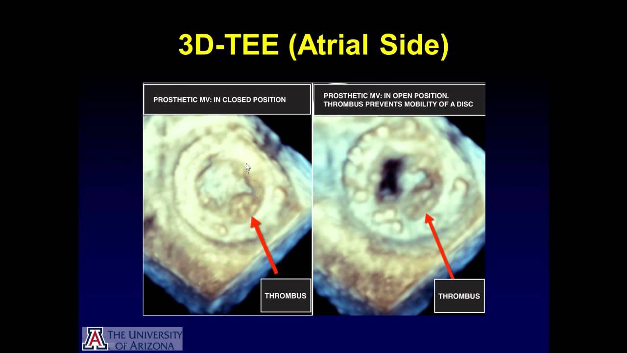
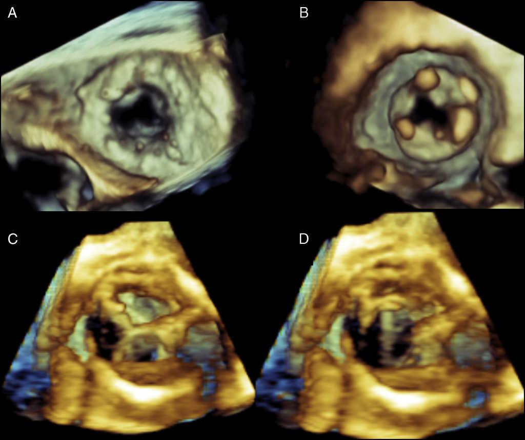
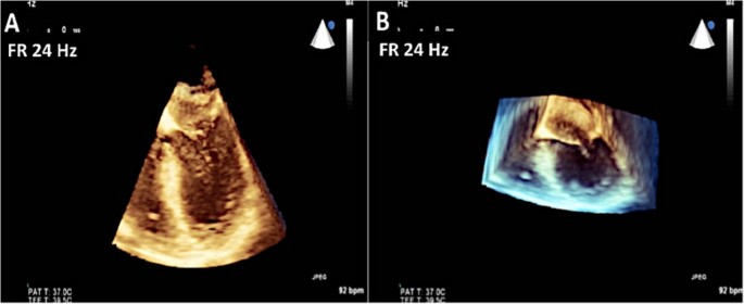
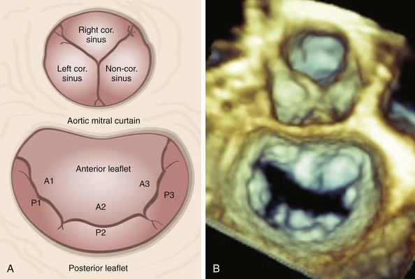
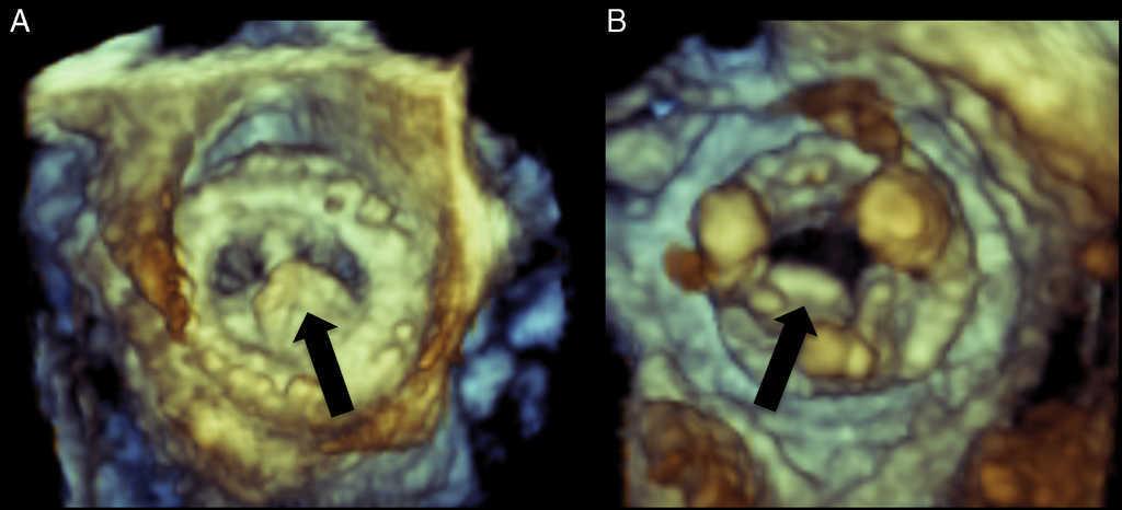
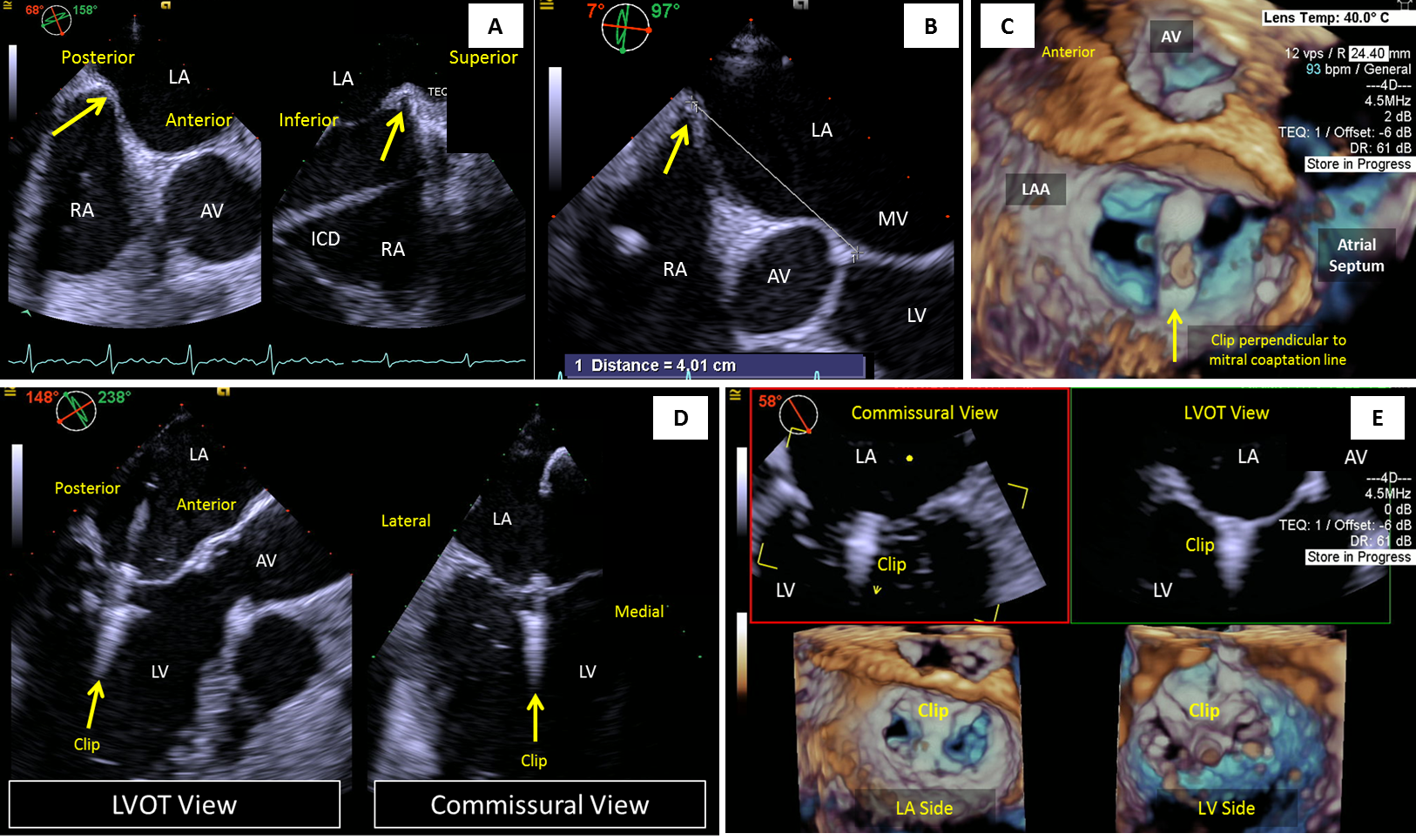
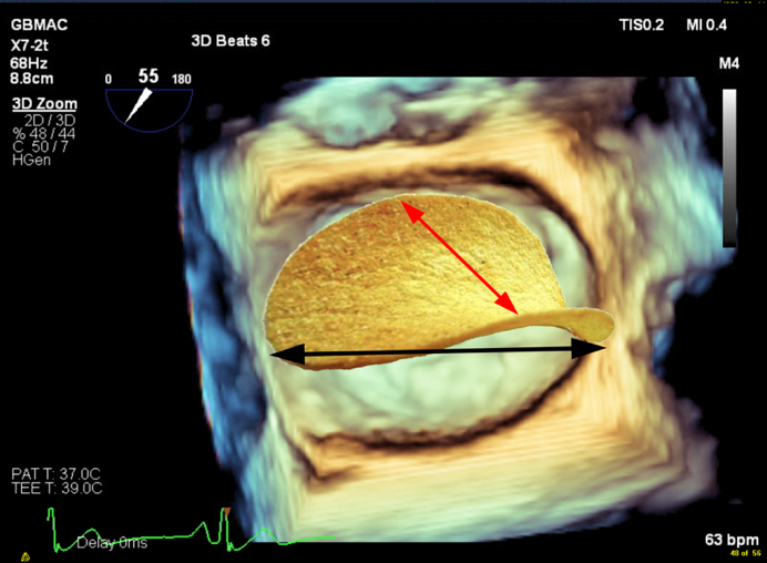
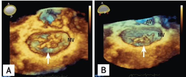
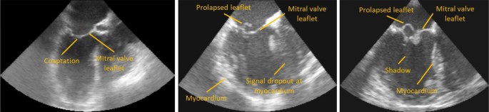
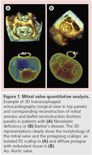
3d tee mitral valve anatomy. The usefulness of ma evaluation using 3d tee imaging has been reported in patients undergoing surgical 22 23 or percutaneous 24 mitral valve repair and in ischemic mr 10 or in the presence of ma disjunction. 3d tee image of the mv with several anteroposterior planes indicated in various colors. 3 d modes such as x plane imaging live 3d imaging 3 d zoom and color 3 d provide incremental information compared to 2 d echo. Assessment of mitral valve anatomy by real time 3 dimensional 3d transesophageal echocardiography tee has proven to be superior compared to 2 dimensional tee 12.
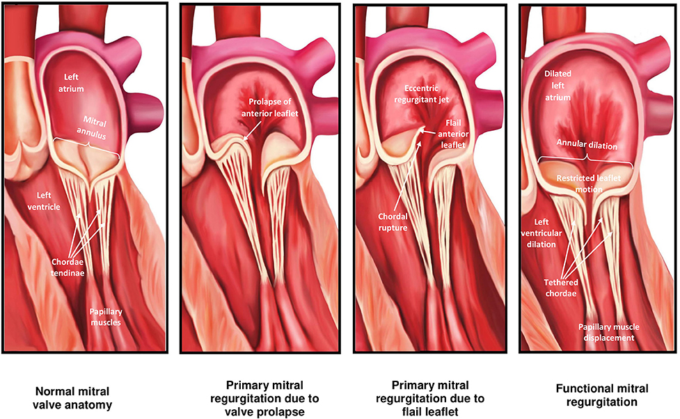
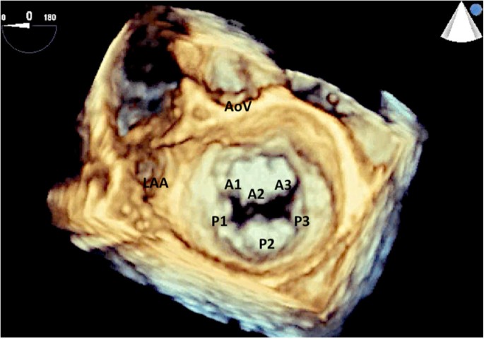
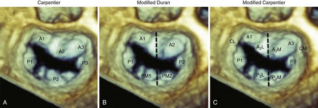
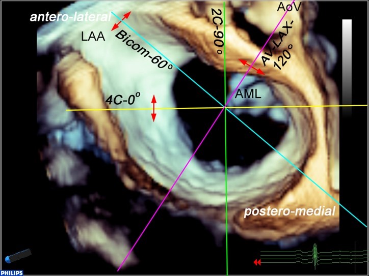
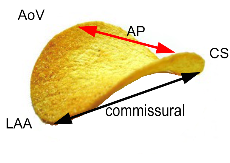
3 d echocardiography is essential for comprehensive evaluation of mitral valve anatomy to assess etiology and mechanism of mitral regurgitation. In complex mitral valve disease anatomy cannot be fully appreciated by only 2 d echocardiography. In 2d each plane it will look like an aortic valve long axis view although the red cut will be imaging the lateral portions a1 p1 and the purple cut will be imaging the medial portions a3 p3 of the valve. 2 and 3 dimensional echocardiography is ideally suited to examine the mitral valve apparatus and has provided insights into the mechanism of mitral valve disease.
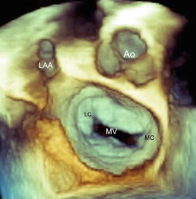
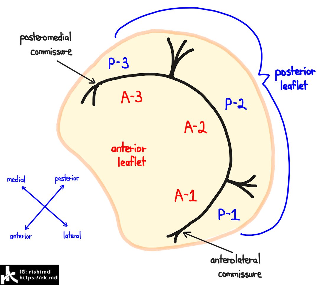
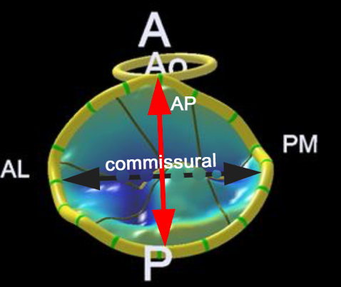
To be a good echocardiographer you will need to be able to relate your echoviews to a specific anatomic location of the mitral valve. Transesophageal 3d echocardiography allows visualization of mitral valve leaflets orifice and submitral apparatus in a manner that is not possible using conventional 2d echo en face views of the mv from atrial and ventricular perspective fully sampled volume not mechanically rotated. Aov aortic valve mv mitral valve. 12 in line with the latter study we found.
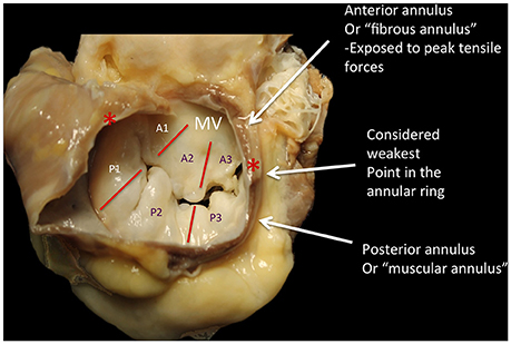
The mitral valve apparatus is a complex threedimensional functional unit that is critical to unidirectional heart pump function. This review details the normal anatomy histology and function of the main mitral valve apparatus components 1 mitral annulus 2 mitral valve leaflets 3 chordae tendineae and 4 papillary muscles. 25 only one prior study showed its usefulness in tmvr sizing for a dedicated prosthesis.

Three Dimensional Echocardiography Is Essential For Intraoperative

Mitral Valve Anatomy Echoboardsacademy

Images Of Note 3d Transesophageal Echo Of The Tricuspid Valve
Https Www Onlinejase Com Article S0894 7317 12 00248 9 Pdf
Frontiers Comparative Anatomy Of Mitral And Tricuspid Valve