How To Do 3d Ultrasound Of Uterus
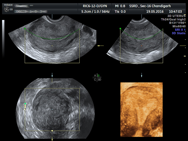
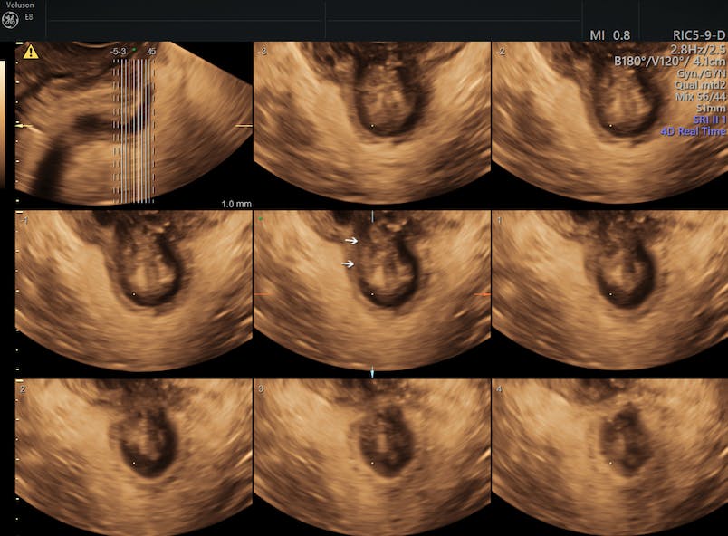
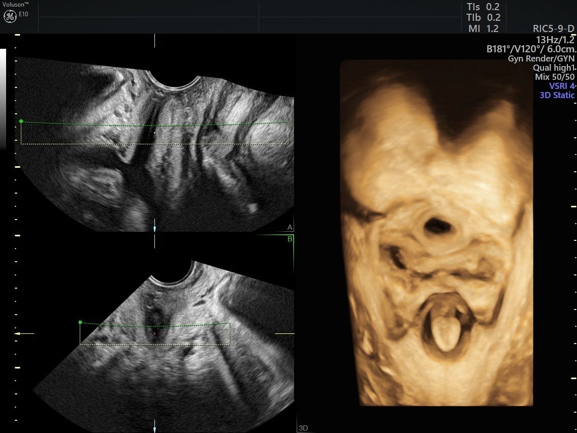
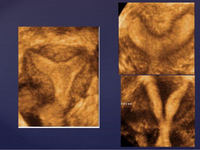
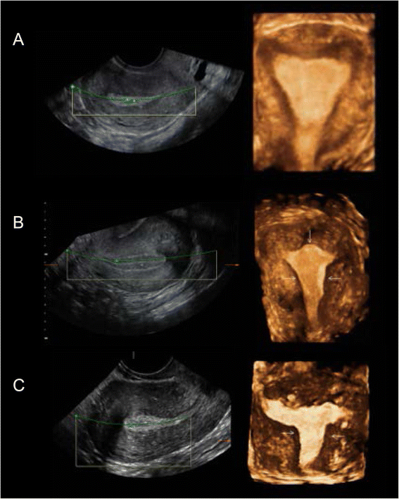
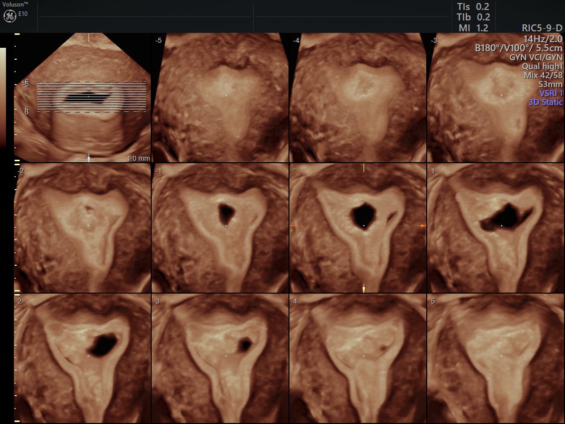
Philips iu22 and epiq philips intellispace portal isp step by step process to obtain midcoronal plane of uterus in volume sonography multiplanar display of a 3d volume 3 orthogonal planes.
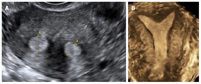
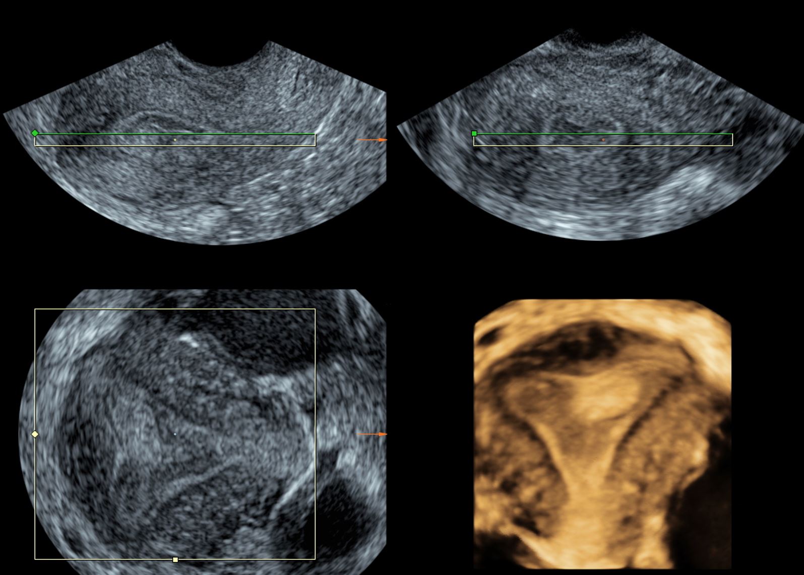
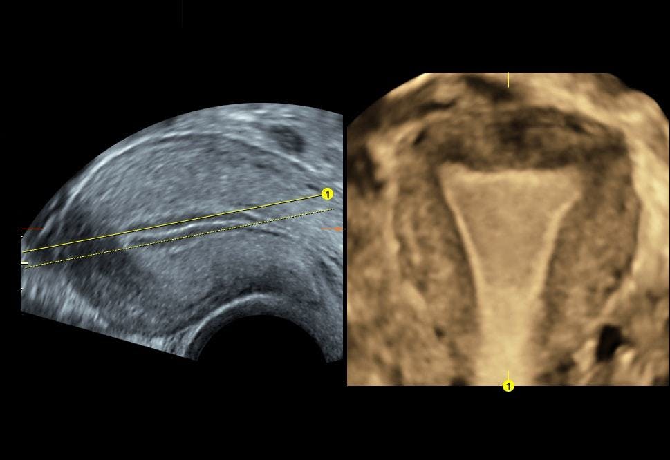
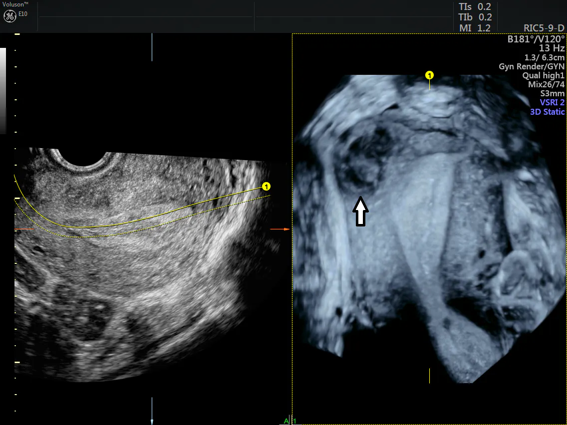
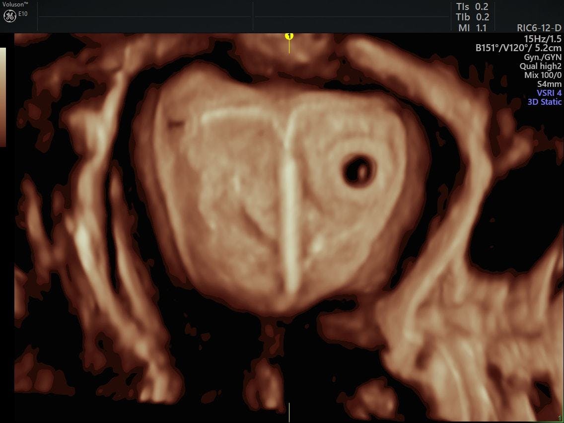
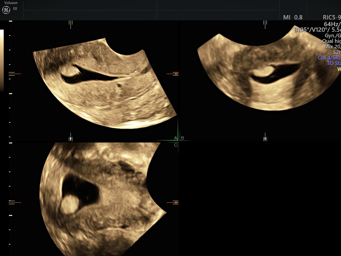
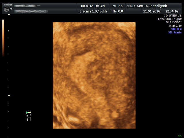
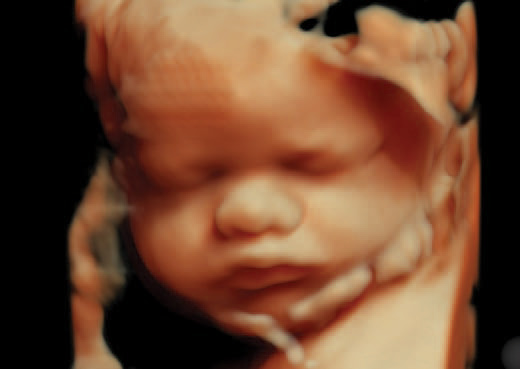
How to do 3d ultrasound of uterus. A sonographer conducts ultrasounds. 3d ultrasound is done using sound waves which helps in capturing clear images of the uterus and the fetus for further study of the pregnancy. Place reference point in mid endometrial stripe ems in sagittal plane use z rotation to align long axis of ems with horizontal put reference point in mid ems in transverse plane apply z rotation on panel c to display uterus in coronal orientation. The application of 3d ultrasound 3dus with increased spatial awareness in the last decade of the 20th century has enabled a detailed assessment of uterine morphology.
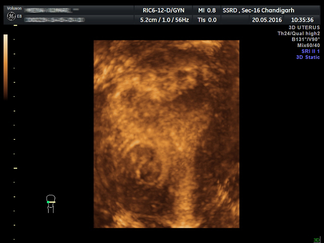
Zoom the image to assess and measure the endometrial thickness. This includes the uterus fallopian tubes. 76376 and 76377 rightthe 3d code is billed in addition to the basic service which in your. A transvaginal ultrasound also called an endovaginal ultrasound is a type of pelvic ultrasound used by doctors to examine female reproductive organs.
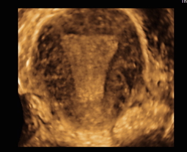
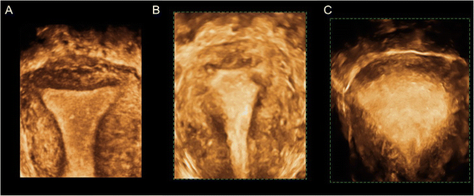
High frequency sound waves are used to create a picture of the uterus. In this plane you should be able to assess the uterus vagina and cervix. The safety of 3d and 4d ultrasounds during pregnancy. Obtaining a 3d coronal image of the uterus.
The 3d ultrasound is generally considered safe as there is no use of radiation insertion of any chemical solutions or use of needles and injections. A small hand held device is lubricated and pressed against the abdomen. Heel the probe to get the bladder over the fundus of the uterus. The freehand technique and automatic acquisition.
Q is there a code for a 3d gyn ultrasound for example to detect endometriosis. Rotate into transverse and angle slightly cranially to be perpendicular to the uterus. Scan sagitally in the midline immediately above the pubis. Or can you only bill the 76856 code ultrasound pelvic nonobstetric b scan andor real time with image documentation.
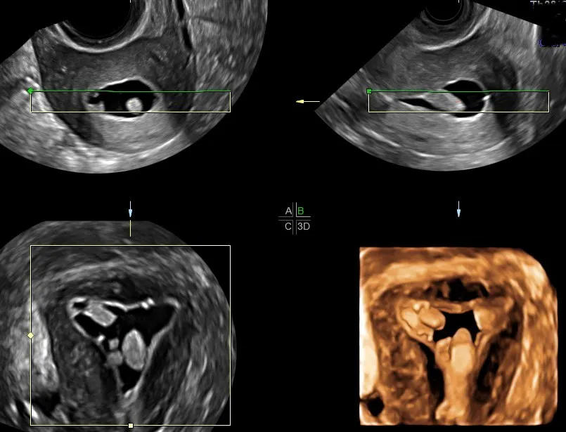
Methods there are two methods for three dimensional volume acquisition. The entire uterine ultrasound procedure usually takes only a few minutes and can be performed in a doctors office in many cases. What is the purpose of 3d ultrasound. A as it happens cpt added new codes this year to allow billing for 3d ultrasound.
Volume acquisition for 3d ultrasound requires specialized ultrasound systems and transducers. A transvaginal compared to the transabdominal approach is generally preferred due to the higher frequency of the probe and the proximity to the pelvic organs which improve image resolutionan adequately enlarged mid sagittal or transverse section of.
1

Assessment Of Pelvic Floor By Three Dimensional Ultrasound In

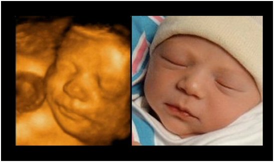
Incredible 3d 4d Scan Of What Unborn Babies Do In The Womb Youtube
Https Encrypted Tbn0 Gstatic Com Images Q Tbn 3aand9gcr0u Lgigl Nr1xj4p0ddxjkcbofapmpgvqeapwegmdtrploe0x Usqp Cau
Saline Infusion Sonogram Diagnosing Endometrial Polyps


/pregnancy-ultra-sound-97530915-588be9f05f9b5874eed3b0d2.jpg)
/GettyImages-157144755-56a773123df78cf772960e9a.jpg)